The Importance of Cytology Testing in Healthcare

A cytology test is a favorable procedure by many doctors because it is non-invasive, accurate, and can effectively identify infections, inflammations, and cancers.
Moreover, the cytology test is considered a powerful diagnosing tool that can detect diseases at early stages leading to improved efficiency of treatment strategy and patient outcome.
Today, we will share updated information about the cytology test, including the preparation process, sample collection methods, the laboratory process, patient experience, and the final results.
Tips for Preparing for a Cytology Test
Exploring Common Cytology Sample Collection Techniques:
The used sample collection method in a cytology test differs based on the examined body area, here are some common methods:
1. Exfoliative Cytology:
This method is usually done by collecting cells that naturally shed from body surfaces, like the cells you might scrape off your skin or those that come from the cervix during a Pap smear.
2. Body Fluid Cytology:
This type works by analyzing in a laboratory the cells found in samples of bodily fluids like urine, sputum, or pleural fluid (the fluid around the lungs), this method is used to diagnose infections and cancers affecting internal organs.
3. Fine-Needle Aspiration (FNA):
The NFA cytology technique provides accurate and fast results, moreover, it involves using a thin, hollow needle to extract cells from solid tissues from a numbed area, like the breast, thyroid, or lymph nodes to investigate lumps or masses.

Understanding the Cytology Laboratory Procedures:
Despite the cytology sample collection method, once the sample is collected, it will be sent to a laboratory for examination, and the cytology procedure in the lab involves several steps:
1- Fixation:
The collected sample is treated with a fixative to reserve the cells, prevent degradation, and guarantee the integrity of the cytology test sample.
Moreover, there are different types of fixatives depending on the type of sample and the required analysis:
- Alcohol-based Fixatives: Usually ethanol and methanol are used to penetrate cells and preserve cellular details quickly, especially Pap smears and other exfoliative cytology samples.
- Formalin: A solution of formaldehyde in water, can perfectly preserve cellular structures, and it is mostly used in the fixation of fine-needle aspiration samples.
- Cytolyte: This technique is mainly used for the fixation of liquid-based cytology, such as urine or body fluids.
- Saccomanno’s Fixative: This fixative contains Carbowax (a polyethylene glycol) and alcohol, and it is mostly useful for sputum cytology, as it helps in preserving respiratory tract cells.
In addition to fixatives, advanced equipment like the LT-YJ2000 Liquid-Based Cytology Smear Processor from Lituo Biotechnology is used to enhance the preparation process, by automating the creation of smears from various cell specimens in just 2.5 minutes based on the membrane filtration method to effectively collect diagnostic materials, ensuring high-quality samples for pathological analysis.
2- Staining:
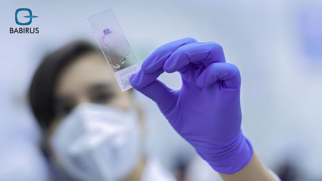
Staining is an important part of the cytology test process, as it allows pathologists to see details more clearly by applying special stains to the sample to highlight cellular structures and make abnormalities more visible under a microscope.
- Papanicolaou (Pap) Stain: Widely used for gynecological and non-gynecological samples, the Pap stain helps separate between normal and abnormal cells, by sharing clear details of the cell’s nucleus and cytoplasm.
- Hematoxylin and Eosin (H&E) Stain: This is a common staining technique in both histology and cytology. Hematoxylin stains cell nuclei blue, while eosin stains the cytoplasm and extracellular matrix pink. This contrast helps diagnosticians easily identify cellular structures and abnormalities.
- Romanowsky Stains: These include Giemsa, Wright, and Diff-Quik stains, which are particularly useful for blood smears and bone marrow samples, by providing detailed visualization of cell types and are often used in hematology.
- Immunocytochemistry Stains: These stains use antibodies to spot specific proteins in cells, and are especially useful for identifying cancerous cells and understanding the cell’s origin, helping in the diagnosis and treatment planning of cancers.
Staining not only enhances the visibility of cellular components but also helps in finding specific cellular markers, as each stain has its particular applications and advantages leading to detect more information about the disease stage and progression.
3-Microscopic Examination:
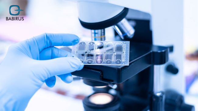
As a main step in the cytology test, the pathologist examines the stained sample under a microscope, looking for any abnormal cells or changes that could point to a specific disease.
Enhancing Patient Experience and Post-Test Care
As we previously mentioned, the cytology test is considered a patient-friendly test, thus, most patients have a positive experience during a cytology test, especially since after the sample is collected, patients can usually resume their normal activities immediately.
However, that does not mean that you should not follow any specific aftercare instructions provided by the healthcare provider, such as monitoring the sample site for signs of infection or other complications.
Even, with the FNA sample collecting method, the worst it could get is some slight discomfort or bruising at the needle site, but this typically resolves quickly.
Interpreting Cytology Test Findings
Cytology testing is preferred by healthcare providers due to its ability to share accurate and vital information about a patient’s health and treatment progress, and the results are usually divided into these categories:
- Negative: This result is comforting for the patients, and is called the safe result indicating that no abnormal cells were detected, indicating no signs of disease, and no action is required.
- Atypical: This result type indicates that some cells appear abnormal, but they are not clearly cancerous, thus, extra testing and diagnostic procedures may be required to determine the cause and explain the findings.
- Positive:Abnormal cells are indicators of cancer or another significant disease, thus, further diagnostic tests or treatments are required.
- Unsatisfactory: The sample could not be properly interpreted.
The importance of understanding cytology results is related to its role in planning the next steps in patient care and treatment strategy, whether it involves additional tests, treatment, or monitoring. Therefore, patients should discuss their cytology test results with their healthcare provider to make informed decisions about their health.
Finally,
All the shared information tells us why the cytology test is a crucial part of modern medical diagnostics, particularly with its valuable insights into a patient’s health with minimal invasiveness and simple steps from preparation to interpreting the results.
Thus, make sure to get access to reliable and advanced medical equipment from a reliable provider like Babirus Medical Equipment and its high-quality tools and solutions.
